Melanoma vs. Normal Mole: How to Tell the Difference
The short answer
Melanoma shows asymmetry, irregular borders, color variation, large diameter, and evolving changes.
Why the Distinction Matters
Melanoma is the deadliest form of skin cancer, responsible for the great majority of skin cancer deaths despite representing only about 1% of skin cancer cases by volume. Early-stage melanoma confined to the skin is highly curable — five-year survival rates for localized melanoma exceed 98%. Metastatic melanoma — spread to lymph nodes or distant organs — carries a dramatically worse prognosis.
The difference between an early diagnosis and a late one often comes down to a patient recognizing a warning sign early and seeking evaluation. This guide provides the clinical framework dermatologists use to distinguish melanoma from a benign melanocytic naevus (mole), translated into language that supports confident self-screening — while being clear about the limits of self-assessment.
Normal Moles: What They Look Like
A benign melanocytic naevus forms from clusters of melanocytes — the pigment-producing cells of the skin — that group together instead of spreading evenly. Virtually everyone has moles; most adults have between 10 and 40. Moles are not dangerous in themselves, though they are the tissue from which some melanomas arise.
Characteristics of a normal, benign mole:
| Feature | Normal Mole |
|---|---|
| Shape | Symmetrical — both halves mirror each other |
| Border | Smooth, even, clearly defined |
| Color | One or two consistent shades of tan, brown, or black |
| Diameter | Usually under 6mm (pencil-eraser width) |
| Stability | Unchanged or changing only very slowly over years |
| Texture | Either flat or uniformly raised (not nodular); surface consistent |
| Sensation | No itching, pain, or bleeding |
Normal moles may be flat or raised, light or dark — variation in these features is normal. What matters is internal consistency and stability over time.
Melanoma: What It Looks Like
Melanoma arises when melanocytes undergo malignant transformation, acquiring DNA mutations — most commonly triggered by UV radiation — that cause uncontrolled, disorganized growth. Because this growth is disorganized, the visual hallmarks of melanoma reflect that chaos: irregular shapes, variable pigmentation, and an inability to maintain stable boundaries.
The ABCDE rule was developed to capture these features in a memorable, teachable framework.
The ABCDE Rule: A Side-by-Side Comparison
A — Asymmetry
| Normal Mole | Melanoma |
|---|---|
| Draw a line through the center in any direction — both halves match | One half noticeably differs from the other in shape, size, or pigmentation |
Asymmetry is the most visually obvious discriminating feature in established melanomas. Even in early lesions, asymmetric growth is one of the first manifestations of the disorganized cell division characteristic of malignancy.
B — Border
| Normal Mole | Melanoma |
|---|---|
| Smooth, even, well-defined edges | Irregular, ragged, notched, blurred, or poorly defined borders |
A melanoma's border may look as if it has been smudged outward, or it may have serrated, scalloped, or jagged projections. The border may appear to fade into the surrounding skin rather than end clearly. Pigment spreading beyond the visible edge of the lesion — into the surrounding normal skin — is a particularly concerning sign.
C — Color
| Normal Mole | Melanoma |
|---|---|
| One or two consistent shades of tan, brown, or black | Multiple colors within the same lesion — may include various shades of brown, black, tan, gray, pink, red, white, or blue |
Color variation within a single lesion is one of the most reliable discriminators. The American Cancer Society notes that concerning lesions may contain "different shades of brown or black, or sometimes with patches of pink, red, white, or blue." DermNet NZ's clinical documentation indicates melanomas may contain up to five or six distinct colors distributed unevenly.
Areas of white or lighter color within a dark lesion may represent regression — areas where the immune system is partially destroying tumor tissue. Pale, white areas within a pigmented lesion are therefore not reassuring; they can indicate an active malignant process.
Areas of red reflect inflammation; blue reflects deeply distributed pigment that scatters light before reaching the surface.
D — Diameter
| Normal Mole | Melanoma |
|---|---|
| Typically under 6mm | Usually larger than 6mm at diagnosis; may be smaller |
The 6mm threshold exists because most melanomas have grown larger than this by the time they present to a dermatologist. However, it is important to understand that diameter alone is neither sensitive nor specific:
- Not all melanomas are large — thin, early melanomas may be well under 6mm. Applying the 6mm rule rigidly can produce false reassurance about small lesions that are nevertheless changing or displaying other ABCDE features.
- Not all large moles are melanomas — some people naturally have benign moles larger than 6mm that have been stable for decades.
A useful clinical refinement: some dermatologists now interpret "D" as "Dark" — a lesion significantly darker than all of the patient's other moles warrants evaluation even if it is under 6mm.
E — Evolving
| Normal Mole | Melanoma |
|---|---|
| Stable over years; if it changes at all, changes are extremely slow | Visibly changing in size, shape, color, or developing new symptoms (bleeding, itching, crusting) over weeks to months |
This is arguably the most actionable criterion. A mole that has been stable for 20 years and suddenly begins to change — even subtly — has lost its reassuring quality. New bleeding, crusting, tenderness, or itching in a previously asymptomatic mole are warning signs that complement visible changes.
Monthly self-examination with a documented baseline (photographs) makes it far easier to detect evolving changes that might otherwise be dismissed as imperceptible.
The Ugly Duckling Sign: The Essential Complement to ABCDE
The ABCDE rule evaluates individual moles in isolation. The ugly duckling sign corrects for this limitation by comparing each mole to the gestalt of the patient's other moles.
Most people develop moles with a broadly consistent "theme" — similar size, color, and shape across most of their moles. A lesion that stands out as dramatically different from all the others — darker, larger, more irregular, lighter, or simply anomalous — is an ugly duckling and should be evaluated even if it does not clearly trigger specific ABCDE criteria.
A PMC-published review proposed formalizing this into the ABCDEF rule by adding "F for Funny looking" to capture the gestalt impression that something looks wrong, even when an explicit criterion cannot be named.
Critical Exception: Nodular Melanoma and the EFG Criteria
Nodular melanoma is one of the most dangerous melanoma subtypes — it accounts for up to 14% of melanomas but a disproportionate share of melanoma deaths. The reason is biological: nodular melanoma grows primarily vertically (into the dermis) rather than radially across the surface. This means it can reach a lethal depth while remaining visually unimpressive by ABCDE standards.
Nodular melanoma may present as:
- A small, symmetrical, dome-shaped nodule
- Uniform dark blue, black, or pink color
- Well-defined or even smooth borders
- Under 6mm — especially early on
- No evolving changes noticed (because it grows rapidly but was not being tracked)
Because ABCDE poorly identifies nodular melanoma, DermNet NZ and other clinical sources recommend the EFG criteria for this subtype:
| EFG Criterion | Feature |
|---|---|
| E — Elevated | A new raised or thickened growth — not flat like a common mole |
| F — Firm | Hard texture when gently palpated; unlike a soft sebaceous cyst or lipoma |
| G — Growing | Rapid growth over days to weeks; stands out from stable background lesions |
A new, firm, raised, fast-growing lesion — regardless of ABCDE status — requires urgent clinical evaluation.
Side-by-Side Summary: Mole vs. Melanoma
| Feature | Benign Mole | Melanoma (superficial spreading) | Nodular Melanoma |
|---|---|---|---|
| Symmetry | Symmetrical | Asymmetrical | Often symmetrical |
| Border | Smooth, defined | Irregular, ragged, notched | May be defined |
| Color | 1–2 consistent shades | Multiple shades; pink, white, or blue | Often uniformly dark |
| Diameter | Usually <6mm | Usually >6mm at diagnosis | May be <6mm |
| Change over time | Stable for years | Slowly changing over months | Rapidly changing over weeks |
| Texture | Flat or uniformly raised | Irregular surface | Elevated, firm nodule |
| Symptoms | None | May develop itching, bleeding | May bleed with minor trauma |
What to Do If You Spot a Concerning Lesion
-
Do not wait. If a lesion concerns you, book a dermatologist appointment promptly. Melanoma is a time-sensitive diagnosis. "Watching it for a few more months" is not a safe strategy for a lesion displaying warning signs.
-
Do not attempt to diagnose it yourself. The ABCDE rule and ugly duckling sign are triage tools, not diagnostic tools. Dermoscopy — examination under polarized light with a dermatoscope — significantly improves clinical diagnostic accuracy over the naked eye, and biopsy with histopathological analysis is the only definitive method for excluding or confirming melanoma.
-
Photograph the lesion and document when you first noticed it and what prompted concern. This history helps the dermatologist contextualize the finding.
-
At the dermatologist's office: If the dermatologist is concerned after clinical and dermoscopic evaluation, a biopsy will be performed — usually an excisional biopsy (the entire lesion removed with a small margin) or a punch biopsy. Histopathological analysis by a dermatopathologist provides the definitive diagnosis.
When a Mole Becomes Higher Risk: Dysplastic Nevi
Some people have moles that are atypical or dysplastic — moles that display some but not all ABCDE features and are considered intermediate between a normal mole and melanoma. Dysplastic nevi are not cancer, but they are a risk marker:
- People with multiple dysplastic nevi have a higher lifetime risk of melanoma
- A single dysplastic nevus in isolation does not dramatically increase risk
- Dysplastic nevi should be monitored; those with features of high-grade atypia on biopsy are often excised with clear margins
The presence of dysplastic nevi warrants closer surveillance and professional monitoring — typically every 6–12 months with whole-body photography.
Common Look-Alikes: Not Every Dark Spot Is a Mole or Melanoma
The differential diagnosis for a pigmented lesion is broad. Conditions that can mimic moles or melanoma include:
- Seborrheic keratoses: Common, benign "stuck-on" growths that are dark, waxy, and warty-appearing; very commonly mistaken for melanoma by patients. They are especially common in people over 50.
- Dermatofibromas: Firm, slightly raised, brownish nodules that often have a subtle dimple when pinched; benign.
- Blue nevi: Benign moles with deep pigment that appears blue-gray on the surface; can be confused with nodular melanoma.
- Hemangiomas: Blood vessel growths that may appear dark red or dark purple; not cancerous.
- Pigmented basal cell carcinoma: In darker skin tones, BCC may appear brown or black and be mistaken for a mole; it requires different treatment.
This is another reason professional evaluation is essential — clinical and dermoscopic assessment by a trained dermatologist is far more accurate than naked-eye assessment of photographs or self-examination alone.
Products that fit: sensitive skin
Picked by code from each product's full ingredient list.

Naturium
Marshmallow Root Barrier Balm
moisturizer · 50 g · 58 ingredients
£21.60−20%Amazon UK
- Fragrance-free
- No essential oils
- No drying alcohol
- No MI/MCI
- No menthol, camphor and witch hazel
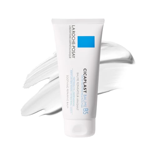
La Roche-Posay
Cicaplast Baume B5 Soothing Repair
moisturizer · 100 ml · 33 ingredients
- Fragrance-free
- No essential oils
- No drying alcohol
- No MI/MCI
- No menthol, camphor and witch hazel

Beauty of Joseon
Red Bean Water Gel
moisturizer · 99.9 ml · 58 ingredients
- Fragrance-free
- No essential oils
- No drying alcohol
- No MI/MCI
- No menthol, camphor and witch hazel

Uriage
Xemose C8+ Lipid-Replenishing Anti-Itch Balm
moisturizer · 500 ml · 24 ingredients
£19.30−17%Amazon UK
- Fragrance-free
- No essential oils
- No drying alcohol
- No MI/MCI
- No menthol, camphor and witch hazel
Amazon UK prices as of 6 November 2026, 06:08 GMT; Amazon sets the final price at checkout.
Frequently asked questions
What does current evidence say about the accuracy of the ABCDE rule in 2026?
The ABCDE rule remains the standard patient education framework endorsed by the AAD and American Cancer Society, but its limitations are well documented in the clinical literature. A key 2015 PMC review noted that applying ABCDE criteria in isolation misses nodular melanoma — which may be small, symmetrical, uniformly dark, and fast-growing without triggering ABCDE. That review, and DermNet NZ's ABCDEFG framework, recommends combining ABCDE with the EFG criteria for nodular melanoma and the ugly duckling sign for a more complete self-assessment. Research into AI-assisted dermoscopy (published in 2024–2025) shows that computational pattern recognition can outperform naked-eye ABCDE assessment, but dermoscopy combined with clinical expertise remains the standard of care, not algorithms alone. The rule is a useful triage tool — not a replacement for professional evaluation.
Can a normal mole turn into melanoma?
Yes, a small proportion of melanomas arise within pre-existing benign moles. Estimates vary — some studies suggest that 20–40% of melanomas arise from or adjacent to an existing naevus, while the majority appear to develop de novo (in normal-appearing skin). This is why monitoring the ABCDE features of existing moles is important: change in a long-stable mole is precisely the 'E' (Evolving) criterion. However, it is equally important not to assume that a long-stable mole is permanently safe — ongoing monitoring is required throughout adulthood.
How do I tell the difference between a mole and a seborrheic keratosis?
Seborrheic keratoses are the most common benign skin growth that patients confuse with melanoma. They typically have a warty, rough, or waxy surface that looks 'pasted on' or 'stuck on' to the skin, often with visible surface crevices. They may be quite dark — dark brown or near-black — and can be quite large, causing alarm. A key distinguishing feature is texture: seborrheic keratoses feel rough and irregular on the surface, whereas melanomas and moles have a smoother texture. Seborrheic keratoses also tend to have a very distinct, sharply defined border with a 'stuck-on' quality rather than emerging from the skin. Despite these distinctions, if you are uncertain, have a dermatologist evaluate it — pigmented BCC can mimic seborrheic keratoses, and there is no benefit to assuming a dark lesion is benign.
Do all melanomas start as a mole?
No. The majority of melanomas — estimated at 60–80% — arise de novo in normal-appearing skin rather than from a pre-existing mole. This is why skin self-examination should look for new lesions, not just changes in existing moles. The appearance of any new dark, irregular, or growing spot on the skin warrants the same ABCDE evaluation as a changing mole. Waiting to see if a new lesion is 'becoming a mole' is not a safe approach.
What happens at a dermatologist's appointment for a suspicious mole?
The dermatologist will first perform a clinical examination — examining the lesion with naked-eye assessment and then with a dermatoscope (a handheld device with polarized light and magnification that visualizes structures beneath the skin surface). Dermoscopy improves diagnostic accuracy for pigmented lesions significantly over naked-eye assessment alone. If the lesion is deemed concerning after dermoscopic evaluation, a biopsy is performed — usually an excisional biopsy (removing the entire lesion with a small margin) for suspicious pigmented lesions, as this provides the complete lesion for histopathological analysis. A dermatopathologist then examines the tissue under a microscope, which is the definitive diagnostic method. Results typically take 1–2 weeks.
Health information, not medical advice.
Beauty Comparison is an independent editorial publication. This page is researched and sourced to the authorities listed below, but it is general information — not a diagnosis or a substitute for personalised advice from a qualified doctor, dermatologist or pharmacist. If your symptoms are severe, spreading, painful, or not improving, see a healthcare professional.Sources
- 01Signs and Symptoms of Melanoma Skin Cancer — American Cancer Society. ABCDE criteria for melanoma: Asymmetry, Border irregularity, Color variation (including shades of pink, red, white, or blue), Diameter >6mm, Evolving changes; normal moles are uniform in color, symmetrical, and stable; the ugly duckling sign; additional warning signs including non-healing sores, pigment spread, and altered sensation.
- 02ABCDEFG of Melanoma — Clinical Criteria — DermNet NZ. ABCDE criteria apply to superficial-spreading melanoma; normal melanocytic naevi are symmetrical with smooth even borders and one or two consistent colors; melanomas may contain up to five or six colors including blue, black, tan, gray, pink, and red; the EFG criteria (Elevated, Firm, Growing rapidly) are required to detect nodular melanoma, which may lack ABCDE features.
- 03The ABCDEF Rule: Combining the 'ABCDE Rule' and the 'Ugly Duckling Sign' — PubMed Central / Journal of Dermatology & Dermatologic Surgery, 2015-01-01. The ABCDE rule in isolation misses lesions that trigger the ugly duckling sign; the proposed ABCDEF framework adds 'F for Funny looking' to combine both screening heuristics; regular self-skin examinations reduce melanoma depth at diagnosis.
- 04Melanoma: Signs, Diagnosis and Treatment — AAD — American Academy of Dermatology (AAD). Melanoma is described by the AAD as 'the most serious skin cancer' because it can spread to other parts of the body; risk factors include UV exposure, family history, and fair skin; dermatologists use dermatoscopes for examination of suspicious spots; early detection significantly improves outcomes.
- 05Advances in the Noninvasive Diagnosis of Melanoma — 40 Years Beyond the ABCDs — PubMed Central / NIH. Dermoscopy significantly improves diagnostic accuracy for pigmented lesions over naked-eye clinical assessment; advances in reflectance confocal microscopy and AI-assisted analysis are complementing but not yet replacing clinical dermoscopy in standard practice.
- 06Melanoma Treatment (PDQ) — Patient Version — National Cancer Institute (NCI / cancer.gov). Invasive melanoma accounts for about 1% of skin cancers but causes the majority of skin cancer deaths; the NCI estimated 104,960 new US melanoma cases and 8,430 deaths in 2025; melanoma staging uses TNM classification with stages 0 through IV based on tumor thickness, ulceration, and spread.