Skin Cancer Warning Signs: The ABCDE Rule Explained
The short answer
Skin cancer warning signs include asymmetry, irregular borders, color variation, diameter over 6mm.
What Is Skin Cancer?
Skin cancer is the uncontrolled growth of abnormal skin cells. It is the most common cancer in the United States — the American Academy of Dermatology (AAD) estimates that one in five Americans will develop skin cancer at some point in their lifetime, and millions of new cases are diagnosed annually. Despite this prevalence, skin cancer is also among the most preventable and, when caught early, among the most treatable cancers.
The three most common types differ fundamentally in origin, behavior, and risk:
| Type | Cell of Origin | Prevalence | Risk of Spread |
|---|---|---|---|
| Basal cell carcinoma (BCC) | Basal cells (deepest epidermal layer) | Most common (~80% of skin cancers) | Very rare metastasis (<1%) |
| Squamous cell carcinoma (SCC) | Squamous cells (middle epidermal layer) | Second most common | Low–moderate; higher in immunocompromised |
| Melanoma | Melanocytes (pigment-producing cells) | ~1% of skin cancers | High — spreads to lymph nodes and organs |
Melanoma is described by the NCI as "a rare form of skin cancer" by percentage, yet it accounts for the vast majority of skin cancer deaths — nearly 20 Americans die of melanoma every day, according to AAD statistics. The NCI estimated 104,960 new melanoma cases and 8,430 deaths in the US in 2025.
The ABCDE Rule: The Primary Self-Screening Framework
The ABCDE rule was developed to give patients and clinicians a structured, memorable framework for evaluating pigmented lesions. Each letter represents a visual feature that distinguishes a potentially cancerous lesion from a benign mole.
A — Asymmetry
Draw an imaginary line through the center of a mole. In a benign mole, both halves should be mirror images of each other. A suspicious lesion has halves that do not match — one half may be larger, differently shaped, or differently pigmented than the other.
Normal moles are typically round or oval and symmetrical. Asymmetry is one of the earliest signs of malignant transformation.
B — Border
A benign mole has smooth, clearly defined, even edges. A suspicious lesion has an irregular, ragged, notched, blurred, or poorly defined border. The edge may appear to bleed into the surrounding skin, or sections of the border may have scalloped indentations.
The irregular border reflects the disorganized, invasive growth pattern of malignant cells.
C — Color
A normal mole is typically one uniform shade — usually tan, brown, or black. A suspicious lesion contains multiple colors within the same lesion — varying shades of brown, black, tan, gray, pink, red, white, or blue.
Color variability results from uneven distribution of melanin, areas of regression (where the immune system is partially destroying the tumor), and inflammation within the lesion. The American Cancer Society notes that concerning lesions may include "different shades of brown or black, or sometimes with patches of pink, red, white, or blue."
D — Diameter
Most benign moles are smaller than 6 millimeters across — approximately the diameter of a pencil eraser. A lesion larger than 6mm warrants evaluation. The AAD notes that melanomas are usually larger than 6mm at the time of diagnosis, though smaller melanomas do exist and diameter alone is not definitive.
Clinical note: Some dermatologists now use "D" to mean "Dark" rather than "Diameter" — this captures the fact that a lesion significantly darker than a patient's other moles, regardless of size, deserves attention.
E — Evolving
Perhaps the most actionable criterion: any mole or lesion that is changing over weeks to months in size, shape, color, or texture is suspicious and should be evaluated. Evolving changes may also include new symptoms — itching, tenderness, bleeding, or crusting in a previously stable mole.
A stable mole may have existed for decades without change. New growth, altered color, or a lesion that begins bleeding spontaneously removes the assumption of stability.
Beyond ABCDE: The Ugly Duckling Sign
The ABCDE rule has one important limitation: it evaluates each mole in isolation. The ugly duckling sign offers a complementary approach — identifying the one lesion that looks strikingly different from all the others on a person's body.
Most people develop moles with a consistent overall pattern. A lesion that looks noticeably different from all of a patient's other moles — the "odd one out" — warrants clinical assessment even if it does not technically trigger ABCDE criteria. A 2015 PMC review proposed combining ABCDE with the ugly duckling sign into a unified "ABCDEF" framework (F for "Funny looking") to capture this gestalt warning without overcomplicating the mnemonic.
The EFG Rule: Recognizing Nodular Melanoma
Nodular melanoma is one of the most aggressive melanoma subtypes, accounting for up to 14% of all melanomas but a disproportionately high share of melanoma deaths — because it grows rapidly vertically (into the dermis) rather than spreading horizontally across the skin surface first.
Critically, nodular melanoma often does not display the standard ABCDE features. It may be small, symmetrical, have defined borders, and be a uniform dark color — and still be lethal.
Dermatologists use the EFG rule to flag nodular melanoma:
- E — Elevated: A raised, dome-shaped, or nodular growth rather than a flat spot
- F — Firm: Hard to the touch, unlike a soft, squishy cyst or lipoma
- G — Growing: Rapid growth over days to weeks rather than the slow change of ABCDE lesions
Any new raised, firm lesion growing rapidly — regardless of ABCDE status — should be evaluated promptly.
Skin Cancer Beyond Moles: Non-Melanoma Warning Signs
Basal cell carcinoma and squamous cell carcinoma — collectively called non-melanoma skin cancers — are far more common than melanoma but present very differently. They do not typically look like moles.
Basal Cell Carcinoma (BCC) Warning Signs
- A pearly or shiny bump with visible blood vessels (telangiectasias) on the surface — the classic nodular BCC appearance
- A flat, scar-like lesion that looks waxy and pale — often the morphoeic (sclerosing) subtype, which is more aggressive
- A pink or red scaly patch that doesn't heal, particularly on the chest or back (superficial BCC)
- A sore that bleeds, crusts, and partially heals, then bleeds again
- In darker skin tones, BCC may appear brown or black rather than pink, which can delay recognition
BCC almost always appears on sun-exposed areas: the head, neck, ears, nose, eyelids, and hands. It is extremely rare for BCC to metastasize, but it can grow deeply, invading cartilage and bone if left untreated.
Squamous Cell Carcinoma (SCC) Warning Signs
- A firm, rough, red or scaly nodule or persistent scaly patch, often in sun-exposed areas
- An open sore or wound that bleeds and doesn't heal within a few weeks
- A wart-like growth that crusts or bleeds
- A raised growth with a central depression that occasionally bleeds
- Lesions on the lip or inside the mouth — oral SCC is less sun-related and may be associated with HPV or tobacco use
Warning Signs That Apply to All Skin Cancers
Beyond the specific type-by-type patterns, the AAD identifies several features that apply broadly to any suspicious skin change:
- Spontaneous bleeding from a lesion — especially one that wasn't injured
- A sore that does not heal within 3–4 weeks
- Pigment spreading from a mole or lesion into the surrounding skin
- Redness or swelling outside the visible border of a mole
- Changed sensation — a previously painless mole that develops itching, tenderness, or pain
- Surface texture changes — oozing, crusting, or scaliness on a previously smooth lesion
Any of these features warrants an appointment with a board-certified dermatologist.
Where Skin Cancer Can Appear: Not Just Sun-Exposed Skin
A common misperception is that skin cancer only develops on areas that receive direct sun exposure. While UV exposure is the primary risk factor for most skin cancers, lesions can develop anywhere on the body, including:
- Scalp and hair-bearing areas (often missed in self-exams)
- Between the toes and on the soles of the feet — acral lentiginous melanoma is more common in people of color and occurs on non-sun-exposed skin
- Under fingernails and toenails — a dark band or streak under a nail (subungual melanoma) may appear as a dark line
- Palms of the hands
- Genitals and perianal skin
- Eyes (ocular melanoma) — appearing as dark spots on the iris
- Mucosal surfaces — mouth, nose, and throat
Subungual melanoma in particular is frequently misidentified as a fungal infection or benign pigmentation. A dark streak extending from the nail bed to the nail edge (the Hutchinson sign — darkening of the skin around the base of the nail) is a warning sign requiring evaluation.
Performing a Monthly Skin Self-Examination
The AAD recommends monthly head-to-toe self-skin examinations. A systematic approach ensures no area is missed:
- Face and scalp: Use a comb to part hair and examine the scalp; use a hand mirror for the back of the head and neck
- Ears: Both the front and back of each ear, including the earlobe
- Trunk and torso: Examine front and back, including the underarms
- Arms and hands: Between the fingers, under and around the fingernails, and the forearms and upper arms
- Genitals: Often overlooked; melanoma can occur here
- Legs and feet: The soles, between the toes, and under and around the toenails
Using two mirrors (a full-length and a hand mirror) or asking a partner to inspect the scalp and back improves coverage. Photograph moles to track changes over time — smartphone photos taken in consistent lighting at the same angle provide a useful baseline.
Risk Factors for Skin Cancer
Understanding risk helps calibrate vigilance. Higher-risk individuals should consider more frequent dermatologist check-ups:
UV Exposure
- History of sunburns, especially blistering sunburns during childhood
- Prolonged or occupational outdoor sun exposure
- History of indoor tanning bed use (tanning beds increase melanoma risk significantly)
- Living at higher altitude or latitudes with intense UV radiation
Personal and Family History
- Previous skin cancer (any type) — significantly increases risk of a subsequent skin cancer
- Family history of melanoma — approximately 10% of melanoma cases occur in people with a family history
- Large numbers of moles (>50) — increases melanoma risk
- Presence of atypical (dysplastic) nevi — moles with irregular features that are not yet cancerous but may be precursors
Skin and Biological Factors
- Fair skin, light hair, and light eyes (Fitzpatrick skin types I and II) — have less melanin to shield UV damage
- A history of easy sunburning rather than tanning
- Age over 50 — particularly for men; melanoma rates in men over 50 increase substantially compared to younger adults
- Immune suppression — organ transplant recipients or people taking immunosuppressive medications are at significantly higher risk
Genetic Conditions
- Gorlin syndrome (basal cell naevus syndrome) — a hereditary condition causing multiple BCCs
- Xeroderma pigmentosum — impaired DNA repair greatly amplifies UV damage
Sun Protection: The Primary Prevention Strategy
UV radiation — from both sun exposure and artificial tanning devices — is the primary modifiable risk factor for all three major skin cancer types. The AAD's prevention strategy:
- Broad-spectrum sunscreen SPF 30+, applied to all exposed skin 15–30 minutes before outdoor exposure, reapplied every 2 hours and after swimming or sweating
- Protective clothing — UPF-rated shirts, wide-brimmed hats, UV-blocking sunglasses
- Shade-seeking during peak UV hours (10am–4pm)
- Avoiding tanning beds entirely — the World Health Organization classifies tanning devices as a Group 1 carcinogen
- Reapplication discipline — sunscreen degrades with UV exposure and sweating; reapplication is not optional
When to See a Dermatologist
The AAD's guidance is unambiguous: see a board-certified dermatologist immediately if you notice anything on your skin that is changing, itching, or bleeding. More specifically, seek evaluation for:
- Any mole or spot that displays one or more ABCDE features
- The ugly duckling — a lesion that looks distinctly different from your other moles
- A raised, firm, fast-growing nodule (EFG criteria for nodular melanoma)
- A sore that bleeds spontaneously or fails to heal within 3–4 weeks
- A dark stripe or band under a fingernail or toenail, particularly with associated skin darkening around the nail base
- A new lesion in a sun-protected area, in someone with darker skin, or in a child
- Any change in a pre-existing mole, even subtle
Annual professional skin exams — during which a dermatologist systematically examines every inch of skin — are recommended for anyone with elevated risk factors. Early detection remains the single most powerful tool for improving skin cancer outcomes: early-stage melanoma is highly curable, while metastatic melanoma remains difficult to treat despite advances in immunotherapy and targeted therapy.
Products that fit: sensitive skin
Picked by code from each product's full ingredient list.

Naturium
Marshmallow Root Barrier Balm
moisturizer · 50 g · 58 ingredients
£21.60−20%Amazon UK
- Fragrance-free
- No essential oils
- No drying alcohol
- No MI/MCI
- No menthol, camphor and witch hazel
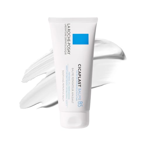
La Roche-Posay
Cicaplast Baume B5 Soothing Repair
moisturizer · 100 ml · 33 ingredients
- Fragrance-free
- No essential oils
- No drying alcohol
- No MI/MCI
- No menthol, camphor and witch hazel

Beauty of Joseon
Red Bean Water Gel
moisturizer · 99.9 ml · 58 ingredients
- Fragrance-free
- No essential oils
- No drying alcohol
- No MI/MCI
- No menthol, camphor and witch hazel

Uriage
Xemose C8+ Lipid-Replenishing Anti-Itch Balm
moisturizer · 500 ml · 24 ingredients
£19.30−17%Amazon UK
- Fragrance-free
- No essential oils
- No drying alcohol
- No MI/MCI
- No menthol, camphor and witch hazel
Amazon UK prices as of 6 November 2026, 06:19 GMT; Amazon sets the final price at checkout.
When to see a professional
See a board-certified dermatologist promptly — within days, not weeks — if you notice any of the following: a mole or spot displaying ABCDE features (asymmetry, irregular border, color variation, diameter >6mm, or evolving changes); a lesion that looks distinctly unlike all your other moles (the ugly duckling sign); a raised, firm, fast-growing nodule (potential nodular melanoma); a sore or wound that does not heal within 3–4 weeks; spontaneous bleeding from a mole; a dark stripe under a fingernail or toenail; or pigment spreading from a lesion into surrounding skin. Do not wait to see if something 'clears up.' Early detection is the single most powerful predictor of a good outcome — early-stage melanoma is highly curable; metastatic melanoma is not. When in doubt, have it evaluated.
Frequently asked questions
Is the ABCDE rule still the recommended standard in 2026?
Yes. The ABCDE rule remains the primary evidence-based patient education framework endorsed by the American Academy of Dermatology and the American Cancer Society. It has been refined and extended — some clinicians now add the EFG criteria (Elevated, Firm, Growing) for nodular melanoma detection and the ugly duckling sign as a complementary tool. A 2015 PMC review also proposed an 'ABCDEF' framework adding 'F for Funny looking' to formally incorporate the ugly duckling concept. The core ABCDE framework, however, remains unchanged and is the standard taught in dermatology worldwide.
Can a skin cancer look normal and not trigger any ABCDE criteria?
Yes — this is particularly true of nodular melanoma, which can be small, dome-shaped, uniformly dark, and have well-defined borders while still being life-threatening. It grows rapidly vertically into the skin rather than spreading outward, making it dangerous but ABCDE-invisible in early stages. Dermatologists use the EFG criteria — Elevated, Firm, Growing rapidly — to flag this subtype. Basal cell carcinoma can also appear as a pale, almost scar-like patch that is easy to dismiss. Any unusual, new, or changing skin finding deserves professional evaluation even if it doesn't check obvious ABCDE boxes.
What is the difference between a mole and a skin cancer?
A benign mole (melanocytic naevus) is a cluster of normal melanocytes that is stable in size, shape, and color over years. It typically has a smooth border, uniform color, and is under 6mm. Skin cancer, particularly melanoma, arises when melanocytes undergo malignant transformation — developing DNA mutations (often UV-induced) that cause uncontrolled, disorganized growth. The ABCDE rule captures the visual consequences of that disorganized growth: asymmetry, border irregularity, color variation, large diameter, and evolving changes. A mole doesn't become a cancer overnight — the transformation is gradual and changes accumulate over months to years.
How common is skin cancer in people with darker skin tones?
Skin cancer is less common in people with darker skin (higher Fitzpatrick types) because increased melanin provides some UV protection, but it is not rare and is frequently diagnosed at later, more dangerous stages. Basal cell carcinoma in darker skin often appears pigmented — brown or black — rather than the classic pearly-pink, leading to misidentification as a benign lesion. Acral lentiginous melanoma, which occurs on palms, soles, and under nails, is disproportionately represented in people of African and Asian descent and is not driven primarily by UV exposure. The common misconception that darker skin is 'immune' to skin cancer contributes to delayed diagnosis and worse outcomes in these populations.
Does tanning bed use really increase skin cancer risk?
Yes, significantly. The World Health Organization's International Agency for Research on Cancer classifies tanning devices as Group 1 carcinogens — the same category as tobacco smoke. Research shows that tanning bed use before age 35 increases melanoma risk by approximately 59%. Indoor tanning delivers intense UV-A and UV-B radiation in concentrated doses without the moderating factors of natural outdoor UV. The AAD strongly recommends against any use of tanning beds or sunlamps.
How often should I see a dermatologist for a skin check?
For individuals with no significant risk factors, an annual professional skin examination is a reasonable baseline. For those with higher risk — a personal or family history of skin cancer, more than 50 moles, a history of blistering sunburns, significant tanning bed use, fair skin, or a history of atypical nevi — more frequent visits (every 6 months) may be recommended. Between professional exams, monthly self-examinations help identify changes early. Supplement with whole-body photography (many dermatology practices offer this) to provide a documented baseline for tracking moles over time.
Health information, not medical advice.
Beauty Comparison is an independent editorial publication. This page is researched and sourced to the authorities listed below, but it is general information — not a diagnosis or a substitute for personalised advice from a qualified doctor, dermatologist or pharmacist. If your symptoms are severe, spreading, painful, or not improving, see a healthcare professional.Sources
- 01Skin Cancer: Signs, Diagnosis and Treatment — American Academy of Dermatology (AAD). Skin cancer is the most common cancer in the United States; one in five Americans will develop it in their lifetime; early detection makes it highly treatable; the AAD recommends evaluation for any changing, itching, or bleeding skin lesion.
- 02Signs and Symptoms of Melanoma Skin Cancer — American Cancer Society. ABCDE rule for melanoma: Asymmetry, Border irregularity, Color variation (including patches of pink, red, white, or blue), Diameter >6mm, Evolving changes; the ugly duckling sign identifies lesions distinctly different from the patient's other spots; additional warning signs include non-healing sores, pigment spreading, and altered sensation.
- 03Melanoma Treatment (PDQ) — Patient Version — National Cancer Institute (NCI / cancer.gov). Invasive melanoma represents about 1% of skin cancers but causes the majority of skin cancer deaths; the NCI estimated 104,960 new melanoma cases and 8,430 deaths in the United States in 2025; higher-risk populations include men over 50 and those with a family history.
- 04The ABCDEF Rule: Combining the 'ABCDE Rule' and the 'Ugly Duckling Sign' in an Effort to Improve Patient Self-Screening Examinations — PubMed Central / Journal of Dermatology & Dermatologic Surgery, 2015-01-01. The ABCDE rule evaluates Asymmetry, Border irregularity, Color variation, Diameter >6mm, and Evolving changes; the complementary ugly duckling sign identifies lesions uniquely different from other nevi; the authors propose combining both into an ABCDEF rule (F = 'Funny looking') to improve patient self-screening coverage.
- 05ABCDEFG of Melanoma — Clinical Criteria Including EFG for Nodular Melanoma — DermNet NZ. Classic ABCDE criteria apply to superficial-spreading melanoma; nodular melanoma requires the EFG criteria (Elevated, Firm, Growing rapidly) because it often lacks ABCDE features; benign moles are symmetrical, stable, and typically one or two colors.
- 06Basal Cell Carcinoma: Symptoms, Causes, and Treatment — DermNet NZ. BCC is caused by DNA mutations in the PTCH tumor suppressor gene, typically triggered by UV radiation; clinical subtypes include nodular (pearly bump with telangiectasias), superficial (scaly patch), and morphoeic (waxy scar-like); dermoscopic features include arborizing vessels and blue-gray ovoid nests; treatment ranges from excision to hedgehog pathway inhibitors for advanced disease.
- 07Comprehensive Insights Into Basal Cell Carcinoma: Causes, Presentation, Prevention, and Modern Therapeutic Approaches — PubMed Central / NIH. BCC represents approximately 80% of all skin cancers worldwide; 3.5 million new US cases annually; UV radiation is the primary cause in fair-skinned individuals; Mohs surgery achieves the highest cure rates (99%); tanning bed use before age 35 increases melanoma risk by approximately 59%; the WHO classifies tanning devices as Group 1 carcinogens.