Malassezia Yeast & Scalp Health: The Fungal Root of Dandruff
The short answer
Malassezia yeast drives dandruff and seborrheic dermatitis by converting scalp sebum into inflammatory free fatty.
What Is Malassezia?
Malassezia is a genus of lipid-dependent (lipophilic) yeasts that form part of the normal human skin microbiome. The genus currently comprises 17 recognized species. Unlike most fungi, Malassezia cannot synthesize their own long-chain fatty acids — the result of genomic losses in fatty acid synthase, Δ9-desaturase, and related enzymes. This dependency on external lipids makes the sebum-rich scalp, face, and upper chest the ideal ecological niches. All healthy humans carry Malassezia on their skin, with colonization beginning at birth and persisting throughout life.
For most people, Malassezia coexists with the host without causing disease. In those who develop dandruff or seborrheic dermatitis (SD), the balance shifts: the yeast's metabolic activity, the sebum environment, and individual immune reactivity together create an inflammatory response that produces the visible scale, itch, and redness of SD.
Why Malassezia Lives on the Scalp
The scalp is one of the sebum-richest sites on the human body, with a high density of large sebaceous glands. Malassezia thrives here because sebum provides exactly the lipid substrates the organism cannot make itself. The organism harvests fatty acids from sebum by secreting a battery of lipases and phospholipases — enzymes that break down triglycerides (the main sebum component) into their constituent fatty acids.
M. globosa is particularly well-adapted for this: its genome contains a high number of lipase-encoding genes and has been identified as the species with the highest lipase activity among all Malassezia species. M. restricta, the other dominant scalp species, also secretes lipases and has been specifically implicated in scalp lipid peroxidation, another inflammatory mechanism.
The Species That Matter for Scalp Health
The Malassezia genus contains 17 species, but for scalp disease, two dominate:
| Species | Role | Key Feature |
|---|---|---|
| M. globosa | Primary driver of dandruff/SD | Highest lipase activity; most prevalent isolate in SD lesions |
| M. restricta | Co-driver of SD; lipoperoxidation | Implicated in squalene peroxide generation on dandruff scalps |
| M. furfur | SD (especially facial), pityriasis versicolor | Most prevalent in European populations in some studies |
| M. sympodialis | Atopic dermatitis (head and neck) | Type I hypersensitivity in atopic individuals |
| M. slooffiae, M. obtusa, others | Minor scalp roles | Present in healthy and diseased scalp; less studied |
A study examining 125 fungal isolates from SD patients found M. globosa (62 isolates) and M. restricta (50 isolates) together accounting for 89.6% of all SD-associated fungi, with both species prevalent on scalp and facial sites alike.
Geographic variation exists: M. restricta and M. globosa predominate in Asian populations, while M. sympodialis is more commonly found in European scalp studies — a difference driven partly by methodology (culture vs. molecular sequencing) and partly by genuine geographic microbiome variation.
How Malassezia Causes Dandruff and Seborrheic Dermatitis
The pathogenic mechanism involves at least three distinct pathways, all of which converge on scalp inflammation.
Pathway 1: Lipase-Driven Free Fatty Acid Generation
This is the best-understood mechanism and the target of most SD treatments:
- Malassezia secretes lipases into the hair follicle and scalp surface
- These lipases hydrolyze sebum triglycerides, releasing free fatty acids (FFAs)
- The organism preferentially consumes saturated FFAs (stearic, palmitic acid) for its own metabolism
- This leaves behind an accumulation of unsaturated FFAs — particularly oleic acid — on the scalp surface
- Oleic acid penetrates the stratum corneum and directly disrupts epidermal barrier function
- Keratinocytes respond to the barrier disruption and the fatty acid itself with pro-inflammatory cytokine release (IL-1α, IL-8, TNF-α)
- The result is the scale, itch, and redness of dandruff and SD
This mechanism explains why Malassezia is able to cause disease without directly "infecting" skin tissue — its metabolic by-products, rather than invasion, drive the inflammatory response.
Pathway 2: Immune Activation Through Pattern Recognition
Malassezia also triggers the innate immune system via multiple receptor pathways:
- TLR2 signalling: Malassezia cell wall components are recognized by Toll-like receptor 2 on keratinocytes and dendritic cells, triggering pro-inflammatory cytokine cascades
- Dectin-2 recognition: The C-type lectin receptor Dectin-2 on dendritic cells recognizes Malassezia mannans, activating downstream CARD9 signalling
- NLRP3 inflammasome: Both M. restricta and M. globosa activate the NLRP3 inflammasome in macrophages and dendritic cells, triggering IL-1β release and caspase-1-dependent inflammation
The adaptive immune response that follows is characterized by Th1, Th17, and Th22 polarization, with elevated IL-17, IFN-γ, and IL-22 in SD lesions. Elevated IL-2 and IFN-γ have been documented in the serum of SD patients compared to healthy controls. This cytokine profile closely resembles psoriasis — which shares histological and treatment overlap with SD.
Pathway 3: Hyphal (Filamentous) Morphology and Virulence
Malassezia normally grows as a yeast (budding single cells), but under certain conditions switches to a hyphal or pseudohyphal filamentous form. This morphological change appears to be pathogenically significant.
A 2022 study examining 90 SD patients and healthy controls found:
- 36.7% of SD patients showed evidence of Malassezia hyphae in skin samples
- No hyphae were detected in any healthy control scalp
- Hyphal presence was positively correlated with clinical SD severity: p = 3.17 × 10⁻¹¹ — an exceptionally strong statistical association
- Following antifungal treatment, hyphal disappearance occurred in parallel with symptom resolution
This finding suggests that the switch from yeast to hyphal morphology represents a shift to a more invasive, tissue-interacting state and may be a key determinant of whether Malassezia causes overt disease in a given individual.
The Paradox: Why Doesn't Everyone Get Dandruff?
Malassezia is ubiquitous — present in essentially every adult scalp — but dandruff and SD affect only 3–12% of the general population. This paradox points clearly to the importance of individual immune reactivity and host factors, not yeast presence alone.
The evidence for an immunological component to SD comes from multiple directions:
- Antifungal response: Treating SD with antifungal agents produces clinical improvement AND significantly reduces Malassezia density — the most direct evidence that the yeast drives the disease
- Immunosuppression amplifies severity: SD affects 34–83% of HIV-positive individuals and 52–59% of Parkinson's disease patients — populations with impaired immune control of commensal organisms
- No consistent load-severity correlation: Some studies find similar Malassezia counts on dandruff-affected and unaffected scalp areas, suggesting that susceptibility — not density alone — determines disease expression
- Specific IgE: Some SD patients show elevated IgE antibodies to Malassezia antigens, suggesting immune sensitization plays a role
The current scientific consensus is that SD represents an abnormal host immune response to a normal commensal, rather than a simple infection with a pathogen. The predisposition appears genetically influenced (family history is a recognized risk factor) and can be amplified by stress, immunosuppression, sebum overproduction, and other triggers.
Sebum and the Scalp Environment: Why Some Scalps Are More Vulnerable
Sebum production is the key environmental variable that determines whether a given scalp is hospitable to Malassezia overgrowth. This explains several clinical observations:
- Age: Sebum production peaks in adolescence (under androgenic hormonal influence) — corresponding to the first major peak in SD prevalence
- Sex: Men produce more sebum than women on average — explaining the consistently higher SD prevalence in males
- Parkinson's disease: Autonomic nervous system dysfunction increases sebum secretion and reduces the facial movements that normally distribute and clear it — providing the concentrated sebum environment in which Malassezia proliferates unchecked
- Scalp sites: The vertex and frontal scalp — highest sebaceous gland density — are the most affected areas
The sebum–Malassezia–inflammation triad is a feedback loop: inflammation from Malassezia metabolites further disrupts the skin barrier, which allows more lipase products to penetrate, sustaining the inflammatory cycle even if yeast numbers temporarily reduce.
Malassezia Beyond the Scalp
While the scalp is the most common site, Malassezia-associated disease occurs across multiple body sites:
| Condition | Body Site | Mechanism |
|---|---|---|
| Seborrheic dermatitis | Scalp, face, chest, ears | Lipase-driven FFA + immune dysregulation |
| Dandruff | Scalp | Milder spectrum of SD without erythema |
| Pityriasis versicolor | Trunk, shoulders | Hyphal invasion of stratum corneum; melanin disruption |
| Malassezia folliculitis | Back, chest, shoulders | Follicular invasion; itch-predominant acneiform eruption |
| Head/neck atopic dermatitis | Neck, cheeks | Type I hypersensitivity; IgE-mediated in atopic individuals |
Dandruff and SD are increasingly understood as "different ends of a disease severity spectrum" — the same process at varying inflammatory intensity, rather than fully distinct conditions. The term seborrheic dermatitis/dandruff complex has been proposed to capture this continuum.
How Malassezia Is Treated
Because Malassezia drives the inflammatory cycle in SD, antifungal treatment is the primary therapeutic strategy. The goal is not to eradicate the organism (impossible, as it is a permanent skin commensal) but to reduce its density and lipase activity to levels the host's immune system can tolerate without triggering inflammation.
First-Line Topical Antifungals
Ketoconazole 2% shampoo is the most studied and best-evidenced treatment. A 2024 narrative review in Cureus (PMC11416180) summarizing multiple clinical trials found:
- 89% of patients treated with ketoconazole experienced improvement or were free of lesions (vs. 44% placebo)
- 73% improvement in dandruff severity score
- Significantly lower recurrence rates vs. zinc pyrithione alternatives
- Protocol: applied twice weekly for 4 weeks for acute treatment; once weekly for maintenance
Ciclopirox 1% shampoo is an effective alternative with a different mechanism (broad-spectrum antifungal + anti-inflammatory). Used three times weekly for 4 weeks in acute treatment.
Selenium sulfide 1–2.5% has direct antifungal and antimitotic effects on scalp keratinocytes. OTC formulations (1%) are widely available.
Zinc pyrithione (in most medicated anti-dandruff shampoos) reduces Malassezia density through a different antifungal mechanism. Effective maintenance option.
Anti-Inflammatory Adjuncts
- Topical corticosteroids: Used short-term (1–3 weeks) during acute flares to reduce inflammation quickly; not suitable for long-term use due to risk of scalp atrophy and telangiectasia
- Calcineurin inhibitors (tacrolimus, pimecrolimus): Preferred for long-term anti-inflammatory maintenance, particularly on the face and ears; do not carry atrophy risk
Systemic Options for Severe or Resistant Cases
For SD not responding to topical treatment, systemic antifungals may be appropriate under dermatologist supervision:
- Itraconazole: 150–200 mg weekly for 2–3 months; 67% complete clearing/marked improvement
- Terbinafine: 250 mg/day for 4–6 weeks; superior to placebo in controlled trials
Emerging Treatments (2025–2026)
A 2025 review (PMC12562114) identified several promising therapies in clinical development:
- Roflumilast 0.3% foam (PDE4 inhibitor): FDA-approved for SD in ages 9+; achieved 79.5% IGA success vs. 58% vehicle control in Phase III trials
- JAK inhibitors (ruxolitinib, delgocitinib): Broad Th1/Th17/Th2 pathway modulation; showing promise particularly for facial SD
- IL-17 and IL-23 inhibitors: Benefit demonstrated in patients with comorbid psoriasis; formal SD approval pending
- Microbiome-targeted approaches: Topical probiotics and postbiotics as experimental adjuncts; evidence still preliminary
The Maintenance Principle
Critically, treating an acute SD flare to clearance is not the end of management. Malassezia repopulates treated scalp within weeks. Maintenance therapy — typically once-weekly antifungal shampoo — is required for most adults to prevent relapse. This is the single most common reason for treatment failure: stopping maintenance after a flare resolves, then experiencing predictable relapse within 4–12 weeks.
For a complete guide to treatment selection and protocols, see Seborrheic Dermatitis on the Scalp and Ketoconazole Shampoo Guide.
The Scalp Microbiome: Malassezia in Context
Malassezia does not exist in isolation on the scalp. The scalp microbiome also includes bacteria (particularly Cutibacterium acnes, Staphylococcus, and Corynebacterium species), which interact with Malassezia in complex ways. The sebum-metabolizing bacterial and fungal communities compete for the same substrates; alterations in the bacterial community can influence Malassezia behavior and vice versa.
This microbiome context explains why antibiotic use can sometimes trigger SD (by reducing bacterial competitors) and why future microbiome-modulating therapies — probiotics, postbiotics — are an active research area. It also explains why anti-dandruff shampoos that only target Malassezia may sometimes be less effective than formulations with both antifungal and antibacterial activity.
Products that fit: itchy, sensitive scalp
Picked by code from each product's full ingredient list.
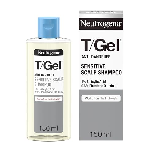
Neutrogena
T/Gel Anti Dandruff Shampoo
shampoo · 150 ml · 15 ingredients
£5.62−13%Amazon UK
- Fragrance-free
- No essential oils
- No MI/MCI
- With piroctone olamine
- With salicylic acid
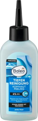
Balea
Professional Scalp Scrub Deep Cleansing
scalp treatment · 150 ml · 10 ingredients
- Fragrance-free
- No essential oils
- No MI/MCI
- With panthenol

OGX
ProGrowth + Peptide Scalp Serum
scalp treatment · 10 ml · 16 ingredients
- Fragrance-free
- No essential oils
- No MI/MCI
- With panthenol

Vanicream
Free & Clear Shampoo
shampoo · 355 ml · 16 ingredients
- Fragrance-free
- No essential oils
- No MI/MCI
- With panthenol
Amazon UK prices as of 6 November 2026, 06:19 GMT; Amazon sets the final price at checkout.
Frequently asked questions
Is Malassezia still considered the primary cause of dandruff in 2026?
The current (2025–2026) scientific consensus views Malassezia as a necessary but not sufficient cause of dandruff and seborrheic dermatitis. The 2025 narrative review (PMC12562114) describes the organism as a 'metabolic and immunologic catalyst rather than a simple commensal.' The evidence strongly supports Malassezia's causal role: antifungal treatments that reduce yeast density consistently produce clinical improvement. However, disease expression requires an abnormal host immune response — which is why most people with Malassezia on their scalp do not develop dandruff. The field has moved from viewing Malassezia as the singular cause to understanding SD as an immune–microbial interaction.
How is Malassezia different from other scalp fungi?
Malassezia is uniquely lipid-dependent — it cannot synthesize its own long-chain fatty acids and must obtain them from the skin's sebum. This makes it exquisitely adapted to sebaceous areas (scalp, face, chest) and means its pathogenicity is mechanistically linked to sebum metabolism. Most other pathogenic scalp fungi (like Trichophyton species in ringworm/tinea capitis) are true dermatophytes that digest keratin for nutrition. Malassezia does not cause tinea capitis or ringworm; its scalp disease is limited to dandruff, seborrheic dermatitis, and folliculitis.
Can you get rid of Malassezia on your scalp permanently?
No — Malassezia is a permanent skin commensal, and achieving long-term eradication is neither possible nor the treatment goal. Antifungal shampoos reduce the yeast population to levels the immune system tolerates without triggering inflammation, but the organism repopulates within weeks of stopping treatment. This is why maintenance therapy (once-weekly antifungal shampoo) is required indefinitely for most people with SD — not to eradicate Malassezia but to keep its density and lipase activity below the threshold that triggers their individual flares.
Does Malassezia cause hair loss?
The relationship between Malassezia and hair loss is indirect rather than direct. Malassezia does not infect hair follicles in the same way dermatophytes do. However, the chronic inflammation of seborrheic dermatitis — including the inflammatory cytokines IL-1α, TNF-α, and IL-17 generated in response to Malassezia — can affect the hair follicle microenvironment. There is growing research interest in a possible link between scalp SD and diffuse hair thinning (telogen effluvium-type shedding), but this is not established causation at the level where clinical recommendations can be made. If you are experiencing significant hair loss alongside SD, discuss both with a dermatologist.
What is the difference between Malassezia folliculitis and seborrheic dermatitis?
Both are caused by Malassezia species, but by different mechanisms and at different anatomical sites. Seborrheic dermatitis involves the surface of the scalp and skin — Malassezia metabolises surface sebum, triggering barrier disruption and immune activation. Malassezia folliculitis involves actual invasion of the pilosebaceous unit (the hair follicle and its attached oil gland), causing follicular inflammation that presents as an itchy, acneiform eruption typically on the back, chest, and shoulders. The two can co-exist; treatment with antifungal agents is common to both.
Is selenium sulfide or ketoconazole better against Malassezia?
Both are effective antifungals against Malassezia, and the choice depends on accessibility, formulation preference, and disease severity. Ketoconazole 2% has the strongest evidence base from controlled trials in SD, with response rates of 73–89% in clinical studies, and superior recurrence prevention vs. zinc pyrithione. Selenium sulfide works by a different mechanism (antifungal + antimitotic on scalp keratinocytes) and is available OTC at 1% in many markets. For mild dandruff, OTC zinc pyrithione or selenium sulfide is appropriate; for true seborrheic dermatitis, ketoconazole 2% is the evidence-based first-line choice, available by prescription in some markets and OTC (1%) in others.
Health information, not medical advice.
Beauty Comparison is an independent editorial publication. This page is researched and sourced to the authorities listed below, but it is general information — not a diagnosis or a substitute for personalised advice from a qualified doctor, dermatologist or pharmacist. If your symptoms are severe, spreading, painful, or not improving, see a healthcare professional.Sources
- 01Seborrheic Dermatitis Revisited: Pathophysiology, Diagnosis, and Emerging Therapies — PubMed Central / NCBI, 2025-01-01. Global SD prevalence 4.38%; M. globosa and M. restricta most implicated; Malassezia acts as metabolic and immunologic catalyst; activates TLR2, Dectin-2, NLRP3 inflammasome; Th1/Th17/Th22 polarization; roflumilast 79.5% IGA success; JAK inhibitors and biologics emerging.
- 02Presence of Malassezia Hyphae Is Correlated with Pathogenesis of Seborrheic Dermatitis — PubMed Central / NCBI, 2022-01-01. Malassezia hyphae detected in 36.7% of SD patients; no hyphae in healthy controls; hyphal presence positively correlated with SD severity (p = 3.17 × 10⁻¹¹); M. globosa (62/125 isolates) and M. restricta (50/125) account for 89.6% of SD fungi; hyphal disappearance parallels antifungal treatment response.
- 03Malassezia-Associated Skin Diseases, the Use of Diagnostics and Treatment — PubMed Central / NCBI, 2020-03-01. Malassezia genus comprises 17 species; colonization begins at birth; M. restricta and M. globosa predominant in Asian populations; M. sympodialis most prevalent in Europe; SD mechanism involves lipase-released oleic acid and arachidonic acid as inflammatory drivers; treatment combines antifungals and anti-inflammatories.
- 04Seborrheic Dermatitis and Malassezia Species: How Are They Related? — PubMed Central / NCBI, 2010-01-01. Malassezia proliferation triggers an immunological response, stimulating inflammation that precipitates SD flares; significant decreases in yeast count after antifungal treatment correlate with visible clinical improvement — the strongest evidence for causation; individual immune predisposition determines whether exposure causes disease.
- 05Seborrheic Dermatitis: Exploring the Complex Interplay with Malassezia — PubMed Central / NCBI, 2025-03-01. Malassezia lipases hydrolyze sebum triglycerides to release oleic acid and other inflammatory FFAs; NLRP3 inflammasome activation releases IL-1β; Dectin-2/CARD9 signalling; Th17 cells and IL-17 are central; immune dysregulation and barrier dysfunction are pathogenically central.
- 06Ketoconazole Shampoo for Seborrheic Dermatitis of the Scalp: A Narrative Review — PubMed Central / Cureus, 2024-08-01. 89% of ketoconazole-treated patients improved vs. 44% placebo; 73% improvement in dandruff severity score; significantly lower recurrence rates vs. zinc pyrithione; 2% ketoconazole twice weekly for 4 weeks is the standard protocol.
- 07An Overview of the Diagnosis and Management of Seborrheic Dermatitis — PubMed Central / NCBI, 2022-08-01. Ketoconazole 2% twice weekly for 4 weeks is first-line; ciclopirox 1% three times weekly is an effective alternative; itraconazole 67% complete/marked improvement; terbinafine superior to placebo; severe/resistant SD warrants HIV screening.
- 08Seborrheic Dermatitis: Causes and Treatment — DermNet — DermNet NZ. SD affects 3–12% of the population; more common in males; Malassezia lipases and phospholipases cleave free fatty acids from sebum; M. furfur, M. globosa, M. restricta are the primary scalp species; SD prevalence elevated in immunosuppressed, HIV, Parkinson's, and neurological conditions.